
Overview
According to a study from Rice university scientists, the dye free molecular atlas of brain of Alzheimer’s patients uncovers a new metabolic change across the brain beyond the amyloid plaque accumulation. It indicates a whole brain metabolic disruption beyond the amyloid protein deposits.

Rice University scientists have compiled a fresh and innovative perspective in the pathological study of Alzheimer’s brain tissues. They have created first comprehensive, label free molecular atlas of the Alzheimer’s brain in an animal model.
All over the world Alzheimer’s claims more lives in the recent decades and its incidence is spiking every year. Till date amyloid plaque deposition is considered as the major cause of the disease and still medical neuro-science has not made great progress in the management of Alzheimer’s and dementia in general.
This study throws light into the fact that metabolic disruption affects brain cells and further research in this regard may help to formulate new strategies to address the onset as well as the progress of Alzheimer’s disease.
Currently, Alzheimer’s is classified among the major diseases causing dementia or loss of memory and eventual progress to nerve degeneration to a wider area of brain incapacitating the patient to lead a normal life on both mental and physical levels.
Previous pathological overview about Alzheimer’s disease
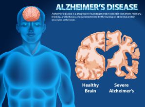
Alzheimer’s disease is a progressive neurodegenerative disorder defined pathologically by the accumulation of extracellular amyloid-beta plaques and intracellular neurofibrillary tangles (NFTs) of hyperphosphorylated tau protein.
Amyloid plaque accumulation happens through the abnormal cleavage of Amyloid Precursor Protein (APP) by beta- and gamma-secretase enzymes. Genetic mutations play key role. Early-onset Alzheimer’s is often caused by mutations in APPPSEN1, or PSEN2 genes. Also, the brain’s inability to remove the amyloid, often due to impaired proteostasis (protein degradation) adds to the pathological change.
Moreover, reduced blood flow, chronic inflammation, and hypoxia (low oxygen) can trigger amyloid plaque production leading to Alzheimer’s disease.
The amyloid proteins disrupt cell-to-cell communication and trigger immune responses (microglial activation). Microglia and astrocytes become activated by protein accumulation, releasing inflammatory cytokines that exacerbate damage.
Oxidative Stress & Mitochondrial Dysfunction exacerbate the crisis. Increased oxidative stress and damage to cellular mitochondria can accelerate neuronal decline.
Tau protein, which normally stabilizes microtubules in neurons, becomes hyperphosphorylated, changes shape, and aggregates into paired helical filaments (tangles). This causes the internal transport system to collapse, leading to neuronal dysfunction and death
Alzheimer’s is is characterized by significant synapse loss and neuron death, primarily affecting the hippocampus and entorhinal cortex, leading to severe brain shrinkage (atrophy).
This study at Rice university gives a deeper outlook towards the emergence and progress of Alzheimer’s disease. Although this study is in the incipient stage and further extensive research is needed to unravel the entire mechanism of metabolic disruption, the study provides new evidence in the pathology of Alzheimer’s disease.
Research method
The scientists of Rice university used an advanced light-based imaging method combined with machine learning, the team examined brain tissue from both healthy and Alzheimer’s affected animals. Their results, published in ACS Applied Materials and Interfaces, reveal that chemical changes linked to Alzheimer’s are not confined to amyloid plaques. Instead, these alterations appear throughout the brain in uneven and complex patterns.
The researchers scanned whole brains slice by slice, compiling thousands of overlapping measurements to build high resolution molecular maps of both healthy and diseased tissue. The imaging process generated large amounts of data, which was analyzed using machine learning. Later they resorted to supervised machine learning, training models to distinguish between Alzheimer’s and non-Alzheimer’s samples. This was a crucial step that aided in determining how different brain regions reflected Alzheimer’s related chemistry.
Research outcome by Rice university
The research team found that the changes caused by Alzheimer’s disease are not spread evenly across the brain. Some areas showed strong chemical changes, while others were less affected. This uneven pattern elucidates clearly about the gradual onset of symptoms in Alzheimer’s disease and why treatments that focus on only one problem have had limited success.
Beyond amyloid plaque accumulation, the study could throw light into the broader metabolic differences between healthy and Alzheimer’s brains.
Levels of cholesterol and glycogen varied across brain regions, with the most dramatic contrasts appearing in areas responsible for memory, specifically in the hippocampus and cortex.
Cholesterol has crucial role in maintaining brain cell structure, and glycogen serves as a local energy reserve.
In a nutshell, this research study offers a more comprehensive view of the disease and opens up new avenues for research both at pathological, investigational and pharmaceutical realms.
Delving deeper into metabolic disruption from other research studies
The findings unveiled include:
- Reduced glucose uptake by brain cells
In alzheimer’s disease significantly reduced cerebral glucose metabolism, particularly in regions like the hippocampus has been detected. Neurons struggle to take up and utilize glucose, leading to a state of “intracellular starvation”.
- Mitochondrial Dysfunction & ATP Failure
- Also, dysfunction in the Tricarboxylic Acid (TCA) cycle, specifically a decrease in ketoglutarate dehydrogenase complex activity.
- Reduced metabolic efficiency causes chronic oxidative stress, which damages brain cells. Microglia cells fail to clear debris and neuroinflammation follows.
- Lipid Metabolism Dysregulation and lipid accumulation which triggers neurodegeneration.
- The Alzheimer’s brain exhibits insulin/IGF resistance, which impairs neuronal glucose transport and worsens energy metabolism.
- Signaling Pathway Disruptions: Dysregulation of the AMPK signaling pathway is closely linked to this impaired energy metabolism.
Conclusion:
I think the new information pertaining to the metabolic dysfunction and oxidative stress may give a new perspective to Alzheimer’s prophylaxis [ prevention], intervention and screening with more metabolic panel analysis.
written by dr sanjana p souparnika
Refernce
- Machine Learning-Enhanced Hyperspectral Raman Imaging for Label-Free Molecular Atlas of Alzheimer’s Brain. ACS Applied Materials, 2025; DOI: 10.1021/acsami.5c22623
- Brain Metabolic Alterations in Alzheimer’s Disease
Carlos G. Ardanaz Department of Pharmacology and Toxicology, University of Navarra, 31008 Pamplona, Spain https://www.mdpi.com/1422-0067/23/7/3785
image courtesy freepik
Leave a Reply